
INTRODUCTION
Oral lichenoid reaction (OLR) or oral lichenoid lesions (OLL) is a term that represent a common end point in response to extrinsic agents (materials, allergens), altered self-antigens, or super antigens. OLL is thus used to describe eruptions of the oral cavity having an identifiable etiology, which are clinically and histologically similar to oral lichen planus (OLP).1
In the literature, different terms are used to refer to these lesions. OLRs have sometimes been considered as part of OLP, and have also been described as contact allergies, oral lichenoid lesions, or contact lesions.2-7 The term OLR was proposed by Finne in 1982 to designate clinically indistinguishable lesions of OLP.2 Oral lichenoid reactions may also result from systemic drug exposure, termed as oral lichenoid drug reaction (OLDR), or local allergic contact hypersensitivity, termed as oral lichenoid contact reaction (OLCR). Oral lichenoid lesions share common clinical and histological features to oral lichen planus (OLP), which is an auto immune disorder exaggerated in the oral cavity.
Since this concept was proposed, these lesions have been described as a response to a wide variety of triggering factors and said to involve several clinical types. OLR presenting as contact-stomatitis, relates to immune mediated hypersensitivity. These have been discovered to be in direct topographic relation with dental restorative materials, most commonly with amalgam. Though OLRs related to non-metallic dental materials are notably less frequent than those to metallic materials, cases presenting the reaction to denture base materials, dental cements, endodontic irrigants have been documented.
Here is a report that presents such an exceptional case of oral soft tissue contact reaction to acrylic base denture material in the form of a soft tissue enlargement occupying the vestibule.
CASE REPORT
A 56 year old female patient, wife of a retired personnel, reported to the Department of Periodontology, Army Dental Centre R & R Delhi Cantt, with the chief complaint of swelling in the upper front gums since the last 3 months and difficulty in wearing dentures due to the same reason. The history of present illness revealed that the patient was asymptomatic 3 months back, and the present complaint started with mild pain in the upper front gums, following which the patient noticed redness in the region. A painless growth appeared in the region which grew to the present size over the past three months.
Upon eliciting the medical history, the patient reported no underlying disease or conditions for which she is seeking, or has undergone any medical consultation. She was not on any medications, no admissions to hospital or a day care facility in the last 6 months, and reported to be otherwise systemically healthy. The patient gave a history of uneventful child bearing and delivery twice, and attainment of menopause at the age of 45 years.
Past dental history revealed multiple tooth extractions over past 20 years. The prime reason for the extractions was dental caries and tooth mobility. The patient had been using dentures for the past 2 years.
Family history was non contributory. On general physical examination she was moderately built, adequately nourished and with erect posture. There were no clinical signs of anemia, cyanosis or icterus. Extra oral examination revealed a bilaterally symmetrical face with no sinus, swelling or scar. A convex lateral facial profile, 5 cm of mouth opening and competent lips at rest were noted. TMJ was normal on palpation and visual examination.
Dental examination revealed completely edentulous maxillary arch, and partially edentulous mandibular arch with presence of the anterior teeth and first premolars bilaterally. The patient wore upper a complete denture and a partial lower denture since last 2 years. On examining the dentures it was noticed that the maxillary dentures were slightly overextended at the labial vestibular area.
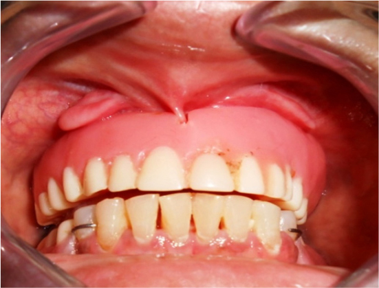
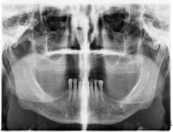
The buccal and labial mucosa was normal in color, contour and consistency. A mucosal overgrowth was observed in the upper anterior vestibular area on either side of the labial frenum (Figure 1). The color of the lesion was pink and in confluence with the adjoining mucosa, the size measured at approximately 2cm in length, 1cm in width, and 0.5cm in breadth. The lesion was pedunculated, non-tender, non-pulsatile, and firm in consistency on palpation. The mucosal attachment was free from the underlying bone and freely movable with the mucosal retraction. Radiographic analysis was carried out using Ortho Pentamo Gram and IOPA radiograph of the involved site. No osseous lesions or pathology was noted (Figure 2).
|
Figure 1: Mucosal overgrowth in the upper anterior vestibular area on either side of the labial frenum.
Click here to view |
|
Figure 2: Ortho Pantamo Gram (OPG) showing no osseous lesions or pathology.
Click here to view |
Based on the patient history, clinical and radio graphical examination a provisional diagnosis of a soft tissue fibroma due to chronic denture irritation was arrived at. A unilateral excisional biopsy of the lesion of the right side, followed by histological examination was planned to arrive at a correct diagnosis. Initially only one lesion was undertaken for biopsy to diagnose the lesion and to avoid additional post operative morbidity and discomfort to the patient.
A complete hematological analysis including complete blood count, INR, APTT, fasting/ post prandial blood glucose estimation was carried out, with normal results.
The surgical site was prepared with extra oral disinfection by 5% povidine iodine scrub, and intraoral 0.2% chlorhexidine rinse. Local anesthesia in the form of ring infiltration with 2% lignocaine HCL with 1:80000 adrenaline was administered by a 30 gauge needle, circumferentially around the lesion in the healthy mucosa.
A 15c blade was used to give a full thickness incision surrounding the lesion on all sides. The incision included 2mm of healthy, uninvolved mucosa on the lesion periphery. The full thickness incision and the involvement of the healthy tissue in the incision design, assured the removal of the lesion in total (Figure 3), failure of which could lead to relapse. The surgical site was sutured with interrupted 3-0 silk, and healing by primary intention was aimed. The patient was advised to discontinue wearing dentures and not to brush on the operated site till the removal of the sutures. Analgesics in the form of Ibuprofen 400mg twice daily for 2 days and mouth rinse with 0.2% chlorhexidine gluconate twice daily for 1 week were prescribed. The patient was recalled after 24 hours to check for any uneventful morbidity, which was ruled out and suture removal was planned 10 days post operatively.

|
Figure 3: A cicumferential full thickness incision and total lesion removal.
Click here to view |
The excised tissue measuring 1.5cm by 1cm (Figure 4) was immediately fixed and transported in 10% formalin solution, to the Department of Pathology, Army Hospital R&R Delhi Cantt for histological examination.

|
Figure 4: Excised tissue measuring about 1.5 x 1cm
Click here to view |
The histological analysis of H & E stained section of the lesion revealed squamous cell epithelial lining showing acanthosis and focal elongation of rete pegs in ’saw tooth’ shaped pattern. Two distinct patterns of plasma cell infiltration (Figure 5) in the dermo-epidermoid junction were seen - One dense and focal and the other scant to moderate diffuse. Dermal edema with no evidence of malignancy were noted. The lesion was identified as a Lichenoid reaction. Based on patient medical and dental history a diagnosis of oral lichenoid contact reaction (OLCR) to dental material, i.e. acrylic based denture material was arrived at.

|
Figure 5: Two distinct patterns of plasma cell infiltration in the dermo-epidermoid junction - One dense and focal (A), and the other scant to moderate diffuse (B).
Click here to view |
The patient was informed and advised to discontinue wearing dentures till the complete healing of the surgical site. Suture removal was done 10 days post operatively. Three month postoperative evaluation revealed satisfactory healing without keloid formation (Figure 6).

|
Figure 6: Three month post-operative evaluation revealed satisfactory healing without keloid formation
Click here to view |
DISCUSSION
The etiology of LP is still under discussion, with a tendency to self-immunity, while the etiology of OLRs is related to the contact with specific agents, such as metallic restorative materials, resins, and drugs, allowing the establishment of a cause-effect relationship.
In the present case, the tissue alteration is thought to be caused by the antigen fixation in the keratinocytes, which are recognized and destructed by cells of the immune system.8, 9 Denture base dental materials play a fundamental role in the appearance of OLCR in the oral mucosa. The reaction to resin materials was reported by Blongren et al.10 Ali et al. studied hypersensitivity to acrylic denture base material, and noted resolution of the lesions of mouth after removal of the prosthesis.11
Lesions of OLCR in relation to dental acrylic prosthesis are located in apposition or in near proximity to the denture base, and lesions are limited to such sites of contact. Typical sites include the lateral borders of the tongue and buccal/ labial mucosa and the buccal/ labial vestibular areas. In the case discussed, the denture flange at the maxillary labial vestibule was evaluated to be a bit over extended. This might have caused repeated impingement of the approximating soft tissue, causing a frank lesion. The lesion with increased vascularity on contact with the denture material might have caused the exuberant lichenoid response as presented.
The lesion was initially thought to be a soft tissue fibroma due to chronic denture irritation. The definitive diagnosis was completely based on the histological analysis of the tissue sample and confirmed the typical lichenoid characteristics of the lesion. Nonappearance of any lesions on other sites of contact with the denture is still not understood. The non-recurrence of the lesion after the excision of the same and advice of discontinuing the wearing of denture is in accordance to the study by Ali et al.11
CONCLUSION
Lichenoid lesions of the oral mucosa present a diagnostic challenge. It may be difficult to distinguish OLP, OLDR, and OLCR on the basis of clinical and/or histological findings. It is essential to obtain a thorough history and perform a complete mucocutaneous examination in addition to specific diagnostic testing (i.e., DIF, IIF, cutaneous patch testing). Generally, OLDR and OLCR resolve once the causative agent has been discontinued.