
Introduction
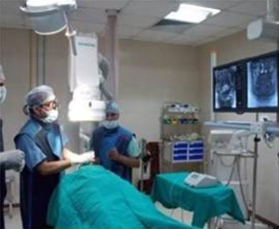
Encouraging results have been achieved using the latest imaging modalities like Dental Cone Beam CT in endodontics but none of these systems can provide a three-dimensional real time imaging. C- arm CT is a dynamic and innovative imaging technique that differs from conventional dental radiography which is static.1 While physicians can observe these live imaging events, dentists have only possibility of monitoring treatment progress is by making radiographs before and after the procedure. As a result, craniofacial surgeries, endodontic treatments and conventional dental implant placement are invariably blind procedures. So, introduction of C-arm systems to endodontic practice will be a giant leap and definitely will provide a cutting edge over existing methods because of its real time imaging advantage. In the present case report, Artis zee C-arm real time imaging system (Siemens AG, Healthcare Sector, Forchheim, Germany) was used for precise detection and location of the separated endodontic instrument as well as to bypass the separated instrument (Figure 1).
|
Figure 1: Endodontic procedure with Artis zee C-arm real time imaging system.
Click here to view |
Case Presentation
A 27 yr old female patient reported with a chief complaint of pain in lower left back tooth. On clinical and radiographic examination, the patient was diagnosed with previously attempted incomplete endodontic treatment and presence of a separated instrument in the middle 1/3rd of mesiobuccal canal of 36. In the present case, use of an operating microscope couldn’t provide the in-depth visualization of separated instrument due to anatomical constrictions of the canal, hence use of C-arm was decided to achieve this. The tooth was isolated using flexi dam (Coltene Whaledent, Switzerland). A preoperative scan was obtained using C-arm system (Figure 2). Access was prepared and ultrasonic tips were used to create troughing around the separated instrument .An attempt to retrieve the separated instrument with instrument retrieval system along with ultrasonic vibration was unsuccessful. So it was decided to bypass the instrument to achieve working length. Bypassing procedure consisted of wedging a # 08 K-file between the canal wall and separated instrument fragment to create a space between them. The file was gradually advanced and withdrawn repeatedly along with use of 17 % EDTA solution to reach the working length. The same was verified by real time visualization on C-arm system for 3 seconds that gave an advantage to observe the file movement, bypassing the separated instrument, eliminating the risk of perforation by the clinician during the procedure. The procedure was repeated using Endosonic files (Dentsply Maillefer, Switzerland) to widen the space created between fragment and canal wall. After widening the space till working length biomechanical preparation was started using # 15/ .04 rotary file (Hyflex CM, Coltene Whaledent, Switzerland) along with 2.5 % sodium hypochlorite irrigation (Figure 3) which was verified by C-arm system for 5 seconds that aided the clinician to observe the movement of rotary file bypassing the separated instrument. Biomechanical preparation was completed using # 25/0.6 rotary files for mesio- buccal and mesio-lingual canal and # 30/0.6 for distal canal (Hyflex CM, Coltene Whaledent, Switzerland). All the three canals were obturated with 6% #25 and 6% #30 single gutta percha cones (Coltene Whaledent, Switzerland) with gutta flow sealer (Coltene Whaledent, Switzerland). C-arm real system was used for real time imaging with an intermittent 3 and 5 second recording mode with the strict adherence to ALARA protocol for both the patient and operator using thyroid collars and lead apron. Post obturation image was obtained using C-arm system (Figure 4).
|
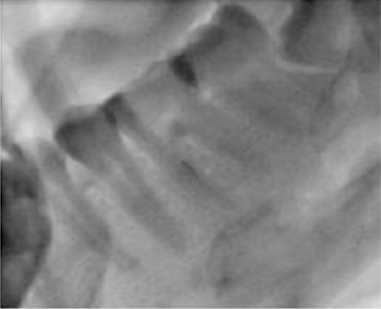
Figure 2: Preoperative Image obtained using C-arm system.
Click here to view |
|
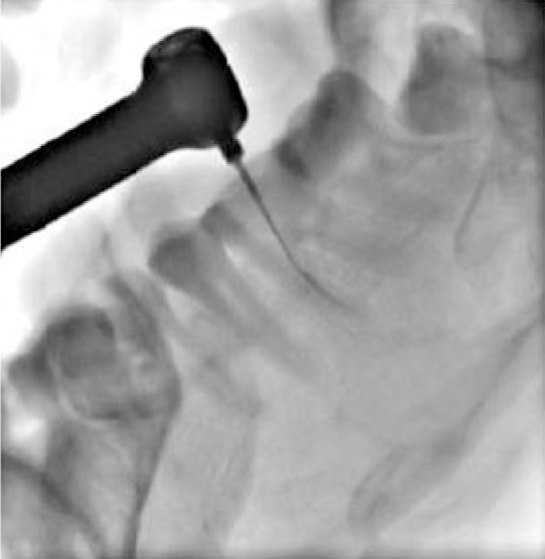
Figure 3: Biomechanica! preparation in Mesiobuccal canal using #15/.04 rotary file.
Click here to view |

|
Figure 4: Post obturation Image obtained using C-arm system.
Click here to view |
Discussion
On literature search in PubMed data base with the words “ C-arm CT in Endodontics” , “C-arm in removal of separated endodontic instrument “ and “ C-arm in bypassing separated endodontic instrument “ no results, “C-arm CT in Dentistry” 4 results, “C-arm Fluoroscopy in Dentistry” 6 results and in Cochrane data base none of the above words yield any results.
In-office C-arm CT has helped to fulfil a growing demand for minimally invasive procedures in traumatology, orthopaedics, endoscopy, paediatrics and urgent care medicine.2 Today, C-arm CT accounts for 35% of the medical imaging market.3
C-arm cone beam computed tomography (CT) is an advanced imaging capability that uses state- of-art C-arm flat-panel fluoroscopy systems to acquire and display three-dimensional (3D) images. C-arm cone-beam CT provides high and low contrast soft tissue (CT-like) images in multiple viewing planes, which constitutes a substantial improvement over conventional single-plannar digital substraction angiography (DSA) and fluoroscopy.4
To obtain 2D radiographic projection data, the C-arm performs a sweep around the patient e.g. over 2000. Up to several hundred images are acquired depending on the acquisition protocol selected. Reconstruction of three-dimensional voxel data sets from 2D raw projection data is performed using a 3D cone-beam reconstruction algorithm. Resulting voxel data sets can be visualized either as cross- sectional images or as 3D data sets using different volume rendering techniques.1, 5-7
The multi-axis stand C arm system facilitates greater flexibility, more accurate, faster movements and better patient coverage, such systems are especially well suited for minimally invasive procedures and surgery.1,8 It offers high-end applications for surgery through real time 3D imaging, high frame rates of 30 frames per second(f/ s), and excellent image quality at low radiation exposure, which is 60-80 % less as compared to Spiral CT.9, 10 As per the manufacturers, the maximum radiation dose for the present system used is 0.25mSv for 5 second exposure time.
In the present case, the real time imaging provided a 3D viewing of involved tooth intricate anatomical details, preventing chances of perforation especially while by-passing the separated instrument thus improving the quality of treatment. The limitation of this system is that it requires complete supine position of the patient and moderately higher exposure protocol and the design that suits a large volume of tissue to be scanned. So, a limited view chair side C-arm system with radiation guidelines is the need of the hour.
Conclusion
Several advanced radiography techniques have been in use in endodontics, namely CT, CBCT, TACT, micro-CT and OCT for in vitro studies. C- arm CT system provides a real time imaging of the root canal system, lesions and surrounding anatomical structures, during clinical procedure, it will definitely gain lot of attention for routine endodontic practice in days to come.